Understanding the assembly mechanism of a virus is critical for the development of antiviral drugs to help combat that virus. Learn more about a collaboration between ESS, Lund University, the University of Copenhagen and the UK's Science & Technology Facility Council's ISIS Neutron & Muon Source to understand the assembly mechanism of hepatitis B virus (HBV).
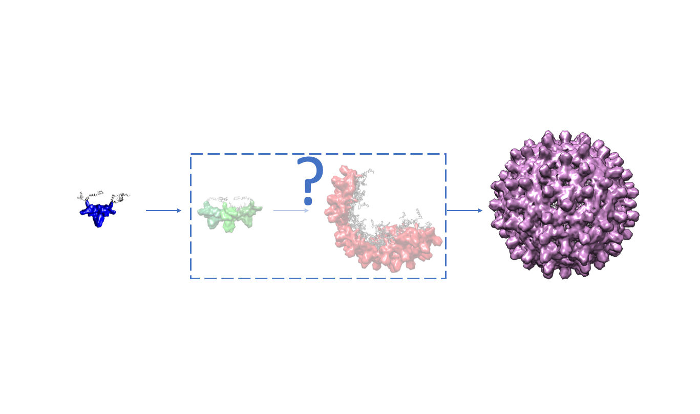
Together with collaborators from Lund University, University of Copenhagen & ISIS, data scientist, Wojciech Potrzebowski, based at ESS Data Management & Software Centre in Denmark, has developed a method that combines molecular modelling with time-resolved small-angle scattering to investigate the self-assembly process of HBV in the presence and absence of ribonucleic acid (RNA).
The understanding of such mechanisms is critical for the development of antiviral drugs, biotechnology, protein engineering and vaccine development. The method was applied to study the HBV virus, but it could be further extended to study other viruses, including the novel coronavirus.
Wojciech is the first co-author of this article leading data analysis aspects. "Together with Ingemar Andre from LU, I wrote and submitted an original proposal to the Swedish Research Council, Vetenskåpradet, or VR, who funded this project, " he explains, adding that he also established a "collaboration with Lise Arleth's group at University of Copenhagen and they offered to share their beam time at ESRF (The European Synchrotron) with us, so that we could perform our measurements. (Lise is a well-established scientist in the field of small angle scattering and she runs a group at Niels Bohr Institute at University of Copenhagen).
"For this study, time-resolved small angle X-ray scattering data was collected at ESRF, which provides sufficient time resolution and quality to observe intermediate structures. It doesn’t, however, provide full insight into RNA/protein composition which can be obtained from experiments using neutrons.
"Once ESS is open to scientists, instruments like ESS's LOKI, with its high flux, will open up for such possibilities in the future," concludes Wojciech.
https://pubs.acs.org/doi/10.1021/acsnano.0c03569