Highlights of Published Papers

Here you will find highlights of scientific papers published by the ESS people. Click on the heading to read the full summary. This list is updated regularly, so be sure to check back for the latest insights.
Science Highlights 2024
The study explores the relationship between shear banding and local concentration fluctuations in dense suspensions of compressible microgels, specifically poly(N-isopropylacrylamide) (PNIPAM)-based microgels. Through a combination of velocimetry, rheology, and small-angle neutron scattering (SANS) performed in the direction of the flow, the authors observed the presence of unsteady, long-lived shear banding at low shear rates. This banding is associated with structural variations and a coupling mechanism between shear and particle migration. The results reveal a central plug-like flow region, corresponding to increases in the local packing fraction and structural correlations in the suspension. The compressibility of the microgels enhances these fluctuations, facilitating particle migration and rearrangement compared to hard-sphere systems.
The findings indicate that shear banding arises from shear-concentration coupling, where gradients in shear rate induce particle migration, altering local packing fractions and structural ordering. A key parameter in this phenomenon is the particle compressibility which allows for particle migration, indeed, even if theoretically predicted such a phenomenon was never experimentally observed for hard incompressible spheres. The study also identifies an upper shear rate limit for observing banding, beyond which uniform flow is re-established. The implications of this research are significant for understanding the rheological behavior of dense suspensions and soft materials, with potential applications in fields like material science and soft matter physics. Further studies are suggested to investigate the role of microgel compressibility and structural deformation in greater detail.
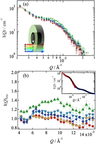
Figure 1: (a) Scattering intensities I(Q) vs Q measured for a shear rate s−1 and for different positions along the velocity profile, as indicated in the sketch. (b) Corresponding excess scattering intensities vs Q. Inset: Scattering intensity I(Q) vs Q in the quiescent state, measured in the shear cell (red circles) and in a normal cuvette (blue squares). Black line: Fit used to extract the excess scattering intensities.
Citation: Bassu, G., Houston, J. E., Lara-Peña, M. A., Kriegs, H., Lettinga, M. P., Porcar, L., Scotti, A. & Laurati, M. Link between permanent shear-banding and local concentration fluctuations in suspensions of compressible microgels. Phys. Fluids 36, 113116 (2024). https://doi.org/10.1063/5.0237526
ESS Contact: Judith Houston
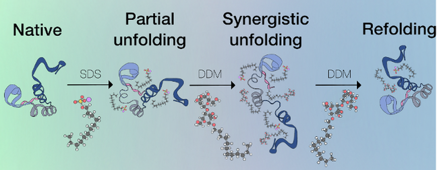
The authors investigate how the combination of an anionic surfactant (sodium dodecyl sulfate, SDS) and a nonionic surfactant (β-dodecylmaltoside, DDM) can influence the unfolding and refolding of proteins. Using three model proteins - human growth hormone (hGH), bovine serum albumin (BSA), and β-lactoglobulin (βLG) - the study demonstrates how different ratios of SDS and DDM affect protein conformational states. The addition of SDS induces protein unfolding, while DDM can partially refold the proteins. However, the process is highly dependent on the specific ratio between the two surfactants. The research highlights that the refolded state does not completely return to the native structure, with some surfactant molecules remaining attached to the protein.
Using various analytical techniques, including fluorescence spectroscopy, circular dichroism (CD), nuclear magnetic resonance (NMR), and small-angle neutron scattering (SANS), the study provides a detailed understanding of the protein-surfactant interactions. The results show that the interplay between SDS and DDM is key to modulating the proteins’ conformational states. The nonionic surfactant DDM can either further destabilize the SDS-unfolded proteins at low concentrations or promote partial refolding at higher ratios. However, full recovery of the native protein structure remains elusive, suggesting that surfactant binding to the protein introduces irreversible changes. These findings offer insights into protein stabilisation mechanisms and the development of formulations in pharmaceutical and industrial applications.
Citation: Hjalte, J., Diehl, C., Leung, A. E., Poon, J. F., Porcar, L., Dalgliesh, R., Sjögren, H., Wahlgren, M. & Sanchez-Fernandez, A. Modulating protein unfolding and refolding via the synergistic association of an anionic and a nonionic surfactant. J. Colloid Interface Sci. 672, 244–255 (2024). https://doi.org/10.1016/j.jcis.2024.05.157.
ESS Contacts: Anna Leung and Jia-Fei Poon
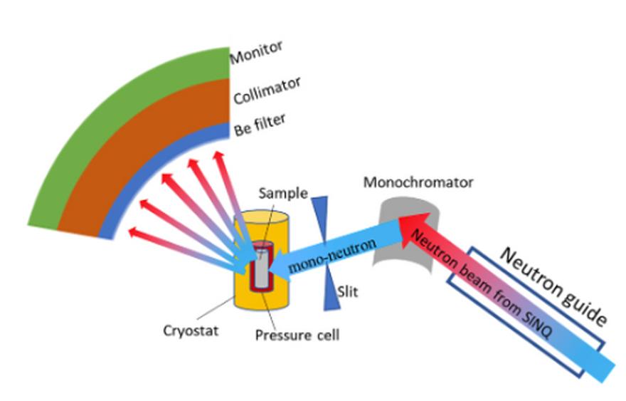
The research addresses the challenges of high background noise in low-temperature high-pressure neutron scattering experiments. The authors identify the main source of background as the piston pressure cell (PC) used in such experiments. Using Monte Carlo simulations, they demonstrate how the pressure cell contributes to significant background noise, which interferes with the detection of signals from small samples. To mitigate this, the study introduces a compact radial collimator (CRC) that can be placed inside the cryostat, close to the sample, to reduce background noise from the sample environment.
The CRC's design and performance are evaluated both through simulations and experimental tests conducted at the CAMEA instrument at the Swiss Neutron Source (SINQ). The experimental results show that the CRC effectively reduces the background noise caused by the pressure cell, particularly at low scattering angles (low-q regions). However, some background noise was introduced by the CRC itself, attributed to its hydrogen-containing polymer material. The paper concludes that using CRCs made from higher concentrations of boron (specifically 10B4C) could further reduce noise, improving the signal-to-noise ratio for future neutron scattering experiments in extreme environments.
Citation: Ma, Z., Lass, J., Mazzone, D. G., Simutis, G., Thürsam, S., Fennell, T., Pomjakushina, E., Bartkowiak, M., Nikitin, S., Bertelsen, M., Willendrup, P., Filges, U., Klauser, C. Sourcing and reducing sample environment background in low-temperature high-pressure neutron scattering experiments. Nucl. Instrum. Methods Phys. Res. A1066, 169634 (2024). https://doi.org/10.1016/j.nima.2024.169634.
ESS Contacts: Mads Bertelsen and Peter Willendrup
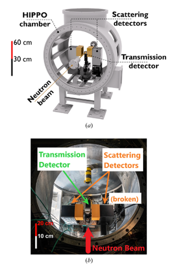
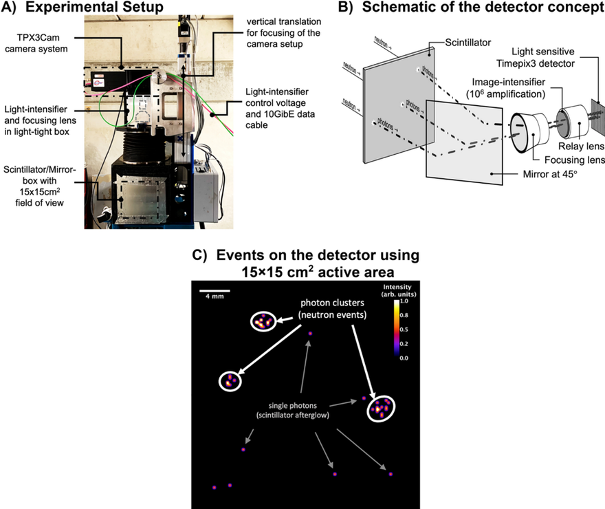
The research highlights the development and demonstration of a novel neutron time-of-flight diffraction system using an event-mode imaging detector based on the Timepix3 technology. This detector system represents a promising alternative to conventional neutron detectors that rely on 3He tubes or neutron-sensitive scintillators. One of the key motivations for developing this new system is the scarcity and cost of 3He gas, which has traditionally been used in neutron diffraction experiments. The event-mode imaging system allows for more flexible and cost-effective data acquisition, and in this study, its performance is benchmarked against conventional systems using a silicon powder diffraction experiment conducted at the HIPPO beamline at Los Alamos National Laboratory.
The results demonstrate that the new imaging detector provides comparable resolution to traditional systems while reducing costs and simplifying hardware requirements. The study shows good agreement between the experimental data, Rietveld analysis, and Monte Carlo simulations using the McStas code, indicating that the system can achieve sufficient resolution for low- to medium-resolution diffraction experiments. Additionally, the authors discuss the potential for future improvements, including increasing the detector's field of view and optimising the scintillator materials used in the system. The demonstration highlights the potential for this new detector technology to be used in a wide range of neutron scattering applications, including diffraction, small-angle scattering, and neutron imaging, with reduced operational and investment costs.
Citation: Jäger, T. T., Losko, A. S., Wolfertz, A., Schmidt, S., Bertelsen, M., Khaplanov, A., Agnew, S. R., Funama, F., Morgano, M., Roth, M., Gochanour, J. R., Long, A. M., Lutterotti, L. & Vogel, S. C. Demonstration of neutron time-of-flight diffraction with an event-mode imaging detector. J. Appl. Cryst. 57, 1107–1114 (2024). https://doi.org/10.1107/S1600576724004448.
ESS Contact: Søren Schmidt and Mads Bertelsen
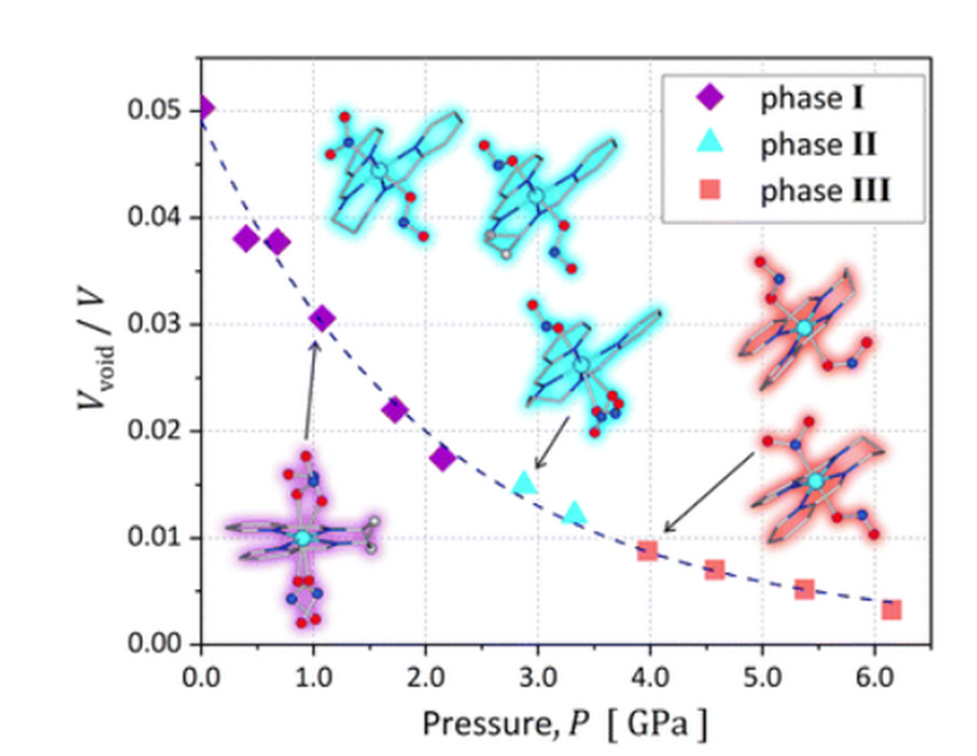
This research focuses on the high-pressure-induced nitrite ligand isomerisation in a nickel(II) complex, accompanied by a piezochromic effect, where the crystal changes colour under mechanical stress. The study presents a novel approach to pressure-induced structural transformations in a rare di-exo-nitrito nickel(II) complex. This system, not photoswitchable, experiences two distinct phase transitions between 0 and 6.2 GPa, resulting in a colour shift from yellow to orange to red. These phase transitions are linked to the isomerisation of nitrite ligands, a process typically induced by light or temperature changes rather than mechanical pressure.
The research offers significant insight into stimuli-responsive materials and their potential applications, such as sensors and optoelectronic switches. By applying high pressure, the researchers demonstrated a unique linkage isomerism in the complex, a phenomenon not previously reported for nitro-to-nitrito transformations. The crystal structure and computational analysis suggest that the exo-nitrito form, though less stable, is strongly supported by the crystal environment, making the isomerisation reversible and efficient under pressure. This study paves the way for further exploration of mechanically induced transformations in molecular materials.
Citation: Potempa, K., Paliwoda, D., Jarzembska, K. N., Kamiński, R., Krówczyński, A., Borowski, P., Hanfland, M. Pressure-induced single-crystal-to-single-crystal nitrite ligand isomerisation accompanied by a piezochromic effect. Chem. Commun. 60, 9194–9197 (2024). DOI: 10.1039/d4cc02898h.
ESS Contact: Damian Paliwoda
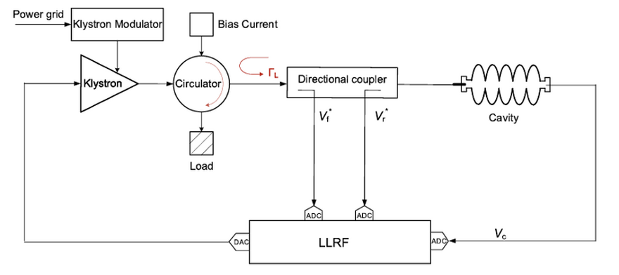
This research focuses on improving the precision of measuring the cavity-loaded quality factor (QL) in superconducting radio frequency (SRF) cavities, specifically addressing challenges related to impedance mismatches. Traditionally, the "field-decay method" has been employed to determine QL, but this method encounters significant errors when there is a mismatch in impedance. To address this, the researchers applied a modified calibration algorithm developed at the China ADS Front End Demo Linac (CAFe), which had been previously validated at the CAFe facility. This study seeks to further validate the effectiveness of the CAFe algorithm at the Test Stand 2 (TS2) facility of the European Spallation Source (ESS) by introducing adjustable impedance mismatches.
The study demonstrates that the CAFe algorithm can accurately measure QL and cavity detuning (Δf) even in the presence of mismatched impedance. By manipulating the circulator bias current at the ESS TS2 facility to vary the reflection coefficient, the researchers introduced impedance mismatches and successfully calibrated QL using the CAFe algorithm. The experimental results show that the proposed algorithm effectively compensates for the errors introduced by impedance mismatches, providing accurate calibration of cavity half-bandwidth and detuning values. This improved accuracy is crucial for ensuring the optimal performance of SRF cavities in large research facilities like the ESS, particularly for applications in high-energy physics and related fields.
Citation: Ma, J., Yang, L., Qiu, F., Wang, M., Zeng, R., Chengye, X., Jingwei, Y., Maiano, C., Goudket, P., Lagoguez, B., Pierini, P. & He, Y. Application of a new cavity-loaded factor calibration algorithm on the Test Stand 2 facility at the European Spallation Source. Radiat. Detect. Technol. Methods 8, 41605-024-00478-5 (2024). https://doi.org/10.1007/s41605-024-00478-5.
ESS Contacts: MuYuan Wang, Rihua Zeng, Cecilia Maiano, Philippe Goudket, Bruno Lagoguez

The authors of this research investigate the behavior of LiFePO4, a magnetoelectric material, in high magnetic fields. The study focuses on understanding the spin-flop transition that occurs when a critical magnetic field of 31 T is applied. Using neutron diffraction in pulsed magnetic fields and electric polarization measurements, the researchers identified the magnetic structure and magnetoelectric (ME) properties in both low and high-field phases. At the critical field, the spin orientation in the material changes from being aligned primarily along the b-axis to reorienting towards the a-axis, suggesting a spin-flop transition. Additionally, the persistence of off-diagonal ME tensor elements above the critical field hints at a more complex magnetic structure than expected.
The study also explores the negligible role of the Dzyaloshinskii-Moriya (DM) interaction in LiFePO4, as no field-induced spin canting was observed. The results were compared with mean-field calculations, which confirmed that the exchange parameters governing LiFePO4's phase diagram fit well with experimental data, supporting the presence of a spin-flop transition. The findings highlight the importance of pulsed-field neutron diffraction and electric polarisation in understanding the magnetic and magnetoelectric behavior of materials like LiFePO4 under extreme conditions. This work contributes to the broader field of multiferroics and magnetoelectric materials, which have potential applications in low-power consumption logic devices and skyrmion-based technologies.
Citation: Holm-Janas, S., Akaki, M., Fogh, E., Kihara, T., Le, M. D., Forino, P. C., Nikitin, S. E., Fennell, T., Painganoor, A., Vaknin, D., Watanabe, M., Christensen, N. B., Nojiri, H., & Toft-Petersen, R. Magnetic structure and magnetoelectric properties of the spin-flop phase in LiFePO4. Phys. Rev. B 109, 174413 (2024). https://doi.org/10.1103/PhysRevB.109.174413.
ESS Contact: Rasmus Toft-Petersen
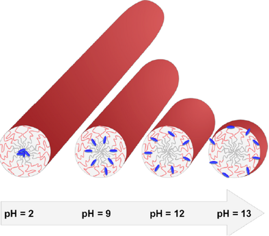
The research presented investigates how the hydrophilicity of an additive, specifically the azo dye "Blue," affects the behaviour of nonionic surfactant micelles formed by C12E5 (pentaethylene glycol monododecyl ether). The study focuses on understanding how changes in the hydrophilicity of Blue, induced by varying pH levels, influence the morphology of the micelles and the localisation of the dye within these structures. The research employs techniques like Small Angle Neutron Scattering (SANS) and 1H NMR to provide insights into micelle structure and behaviour as hydrophilicity shifts from more hydrophobic (protonated BlueH) to highly hydrophilic (bivalent anion Blue2−).
The results reveal that increasing the hydrophilicity of the dye (by increasing pH) leads to notable changes in micelle morphology, transitioning from elongated, rod-like structures to more spherical forms. Additionally, the localisation of the dye within the micelles shifts as its hydrophilicity changes, with more hydrophilic forms of Blue residing closer to the outer regions of the micelle, while more hydrophobic forms penetrate deeper into the micellar core. The study highlights how additive hydrophilicity can significantly influence the structural and phase behaviour of surfactant systems, offering potential applications in fields like dye solubilisation and delivery systems.
Citation: Müller, W., Sroka, W., Schweins, R., Nöcker, B., Poon, J.-F., & Huber, K. Impact of additive hydrophilicity on mixed dye-nonionic surfactant micelles: micelle morphology and dye localization. Langmuir 40, 8872–8885 (2024). https://doi.org/10.1021/acs.langmuir.4c00012.
ESS Contact: Jia-Fei Poon
The authors of this research report the structural and magnetic changes that occur in magnetite (Fe3O4) nanoparticles during chemical lithiation. The research focuses on nanoparticles as potential anode materials for lithium-ion batteries due to their high theoretical energy storage capacity. The study uses various techniques, including X-ray diffraction, electron microscopy, and magnetic measurements, to explore the effects of lithiation at different levels (x = 0, 0.5, 1, and 1.5) on the structure and magnetism of 70 nm cubic-shaped Fe3O4 nanoparticles.
The findings reveal that as lithium is introduced, the magnetite nanoparticles undergo a structural transformation from the spinel phase to a rock salt phase at higher lithium concentrations. This transformation begins when lithium occupies the octahedral sites in the spinel structure and displaces iron ions, leading to a reorganization of the crystal lattice. Magnetization measurements indicate that lithiation results in the formation of antiferromagnetic LiFeO2 subdomains, which increase the interfacial exchange coupling, contributing to a change in magnetic properties. These insights highlight the interplay between structural changes and magnetic behaviour in lithiated Fe3O4, advancing the understanding of their performance in lithium-ion batteries.
Citation: Ulusoy, S., Feygenson, M., Thersleff, T., Uusimäki, T., Valvo, M., Roca, A. G., Nogués, J., Svedlindh, P. & Salazar-Alvarez, G. Elucidating the lithiation process in Fe3−δO4 nanoparticles by correlating magnetic and structural properties. ACS Appl. Mater. Interfaces 16, 1234–1245 (2024).
ESS Contact: Mikhail Feygenson
Ever wondered what’s hiding inside a meteorite or the aftermath of an ancient asteroid impact? Thanks to cutting-edge technology, scientists are now delving deep into these cosmic relics without even scratching their surfaces!
With upcoming sample-return missions, such as the Mars Sample Return Campaign, the need for non-destructive methods to maximise scientific output is crucial. The recent paper published in JGR Planets delves into the application of neutron and X-ray tomography, exploring and reviewing its effectiveness.
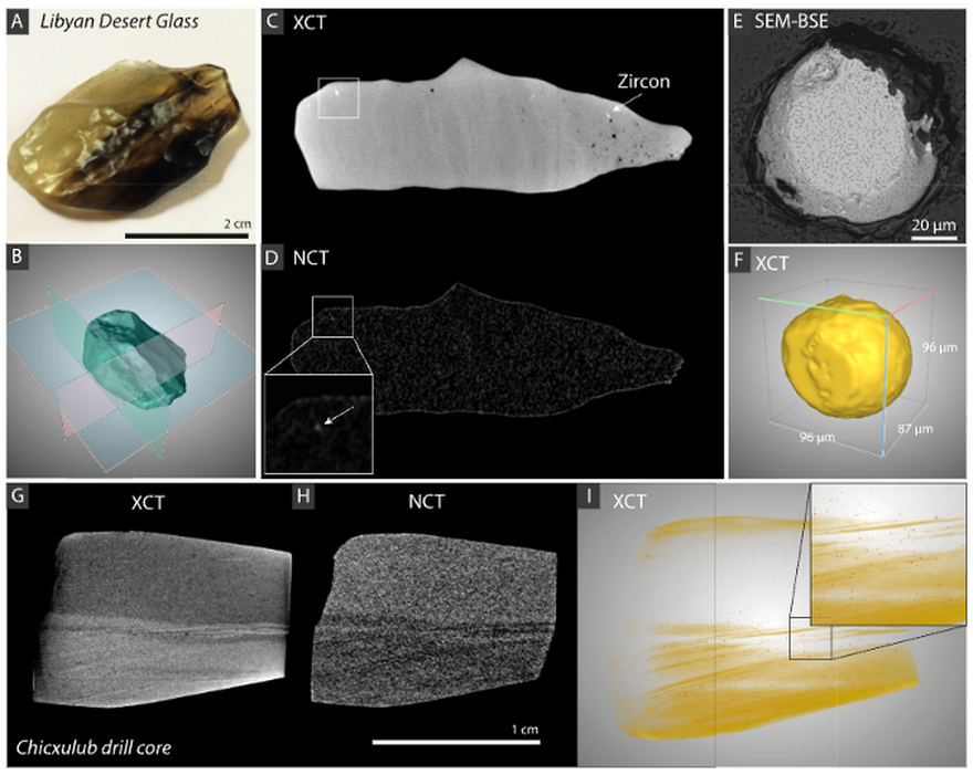
The study presents four case studies involving Libyan Desert Glass, Chicxulub impact core samples, Luizi impact melt rock, and a Martian meteorite (MIL 03346). By combining these tomography techniques, the researchers revealed hidden features such as projectile material and hydrous phases, providing insights into impact cratering processes and aqueous alteration in extraterrestrial samples.
Neutron and X-ray computed tomography (NCT and XCT) are complementary techniques; neutrons interact with atomic nuclei, while X-rays interact with electrons. This allows the two methods to highlight different materials within samples. In this study, the authors successfully identified meteoritic material in Libyan Desert Glass and hydrated phases in the Luizi impact melt rock and the Martian meteorite.
These findings illustrate the potential of combined tomography methods for future planetary exploration. By enabling detailed analyses without contamination, these techniques represent a powerful approach for studying precious samples, shedding light on the history of our solar system.
Citation: Martell, J., Alwmark, C., Woracek, R., Alwmark, S., Hall, S., Ferrière, L., Daly, L., Bender Koch, C., Hektor, J., Johansson, S., Helfen, L., Tengattini, A. & Mannes, D. Combined Neutron and X-ray Tomography—A Versatile and Non-Destructive Tool in Planetary Geosciences. J. Geophys. Res. Planets 129, e2023JE008222 (2024).
ESS Contact: Robin Woracek
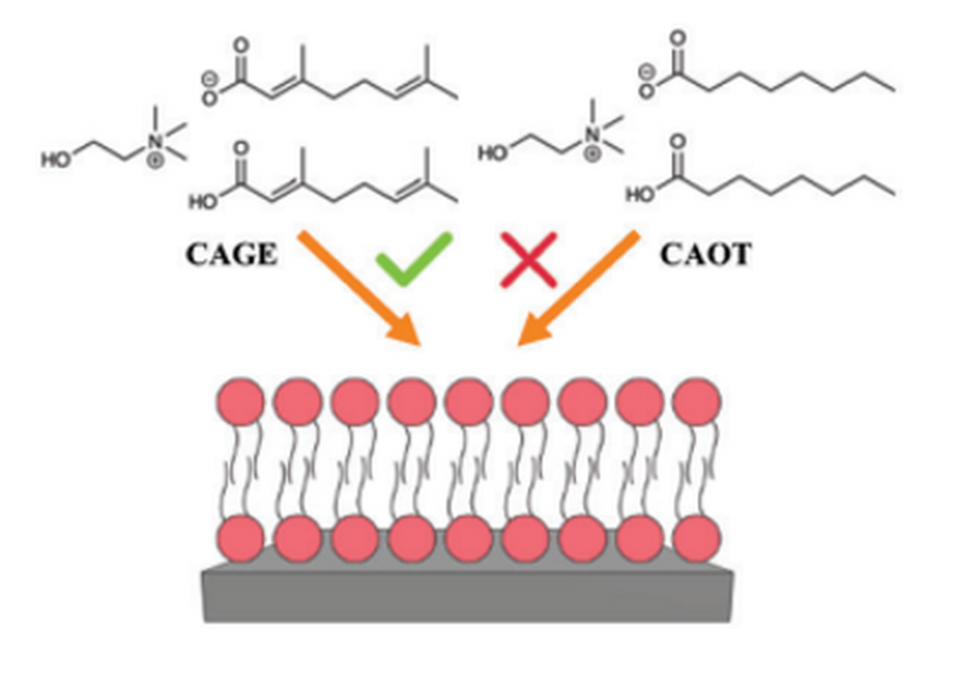
This research aims to investigate how two deep eutectic solvents (DES), CAGE and CAOT, interact with lipid bilayers. CAGE, a mixture of choline and geranic acid, has been shown to facilitate transdermal delivery of large molecules like insulin. The study aims to explore its mechanism by comparing it to CAOT, a related DES. Using neutron reflectivity, the researchers studied six systems involving phospholipid bilayers and discovered that CAGE mildly disrupts bilayers by inserting into and diffusing across them while maintaining structural integrity. In contrast, CAOT showed greater disruption, causing swelling and the formation of solvent patches.
The study reveals that the anionic components of these solvents play a key role in their interaction with lipid membranes. CAGE increases hydration in the bilayer without significant lipid removal, which is crucial for its transdermal delivery capabilities. CAOT, however, disrupts the bilayer more aggressively, exchanging and removing lipid molecules. The research provides insights into how these DESs interact with membranes, laying the groundwork for optimising them for pharmaceutical applications, particularly in enhancing drug delivery through the skin.
Citation: G.M. Neville, A.-M. Dobre, G.J. Smith, S.,Micciulla, N.J. Brooks, T. Arnold, T. Welton, K.J. Edler; Interactions of Choline and Geranate (CAGE) and Choline Octanoate (CAOT) Deep Eutectic Solvents with Lipid Bilayers. Advanced Functional Materials, 34(2), doi: 10.1002/adfm.202306644
ESS Contact: Tom Arnold
Science Highlights 2023
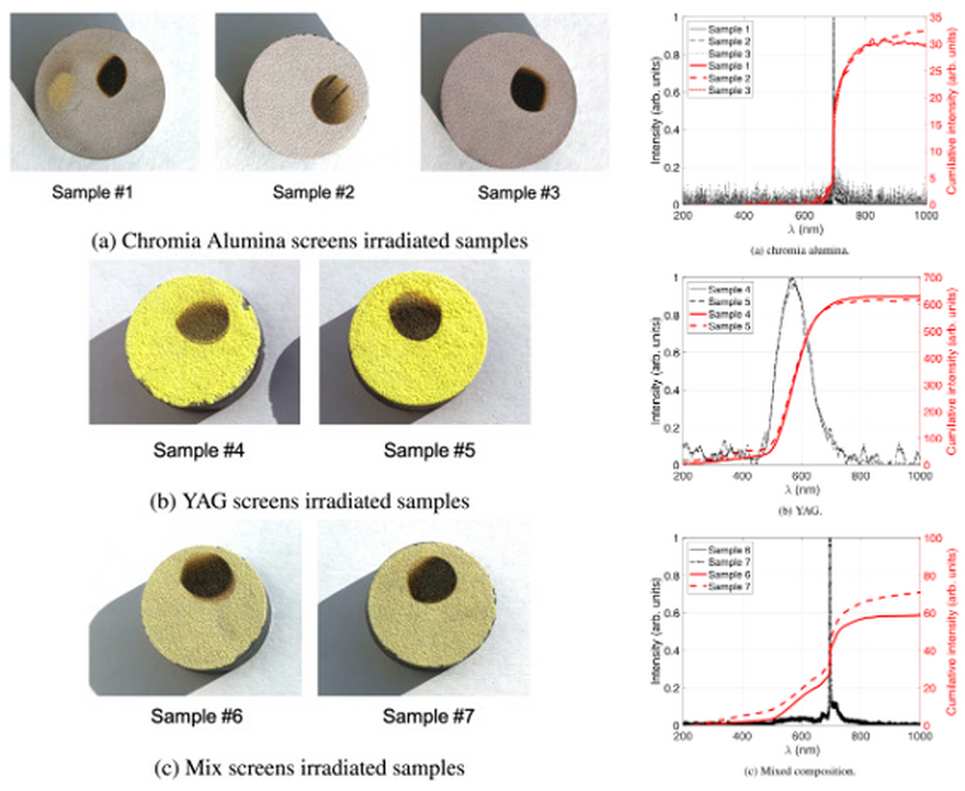
The European Spallation Source (ESS) will deliver a pulsed 2 GeV proton beam with an average power of 5 MW to its tungsten target, each pulse being raster-scanned on the target wheel. Controlling the power-density distribution and position of the proton beam as it hits the target wheel is crucial to limit the damage to the target wheel and surrounding infrastructure. Luminescent coatings are used as scintillating screens to measure the beam parameters such as position and density distribution. Chosen luminescent coatings must have sufficient luminescence yield even at elevated temperatures and be radiation-hard.
This work is part of an ongoing project to qualify suitable luminescent screens. The materials investigated were chromia alumina (Al2O3:Cr), YAG (Y3Al5O12:Ce) and a mixture of the two. The screens, fabricated by flame-spraying scintillating materials onto stainless steel sample chips, were characterised by evaluating their luminescent yield (photons per MeV), their radiation resistance and their spectral response under irradiation with a proton beam at 2.55 MeV. Several key findings are reported including that high temperature YAG (>200°C) retains higher luminescence yield than chromia alumina at elevated temperature which is a huge advantage. There are also signs that the mixed composition screens may permit absolute temperature measurement – a very useful additional property.
Citation: Frost, R. J. W., Thomas, C. A., Elfman, M., Johansson, R., Hartl, M., Kocevar, H., Michel, K., Joshi, S. & Bjorklund, S. Preliminary results from a study of luminescent materials-For application in the beam imaging system at the ESS. Nucl. Instrum. Methods Phys. Res. Sect. B-Beam Interact. Mater. Atoms 540, 227-233, doi:10.1016/j.nimb.2023.04.015 (2023).
ESS Contacts: Cyrille Thomas / Monika Hartl
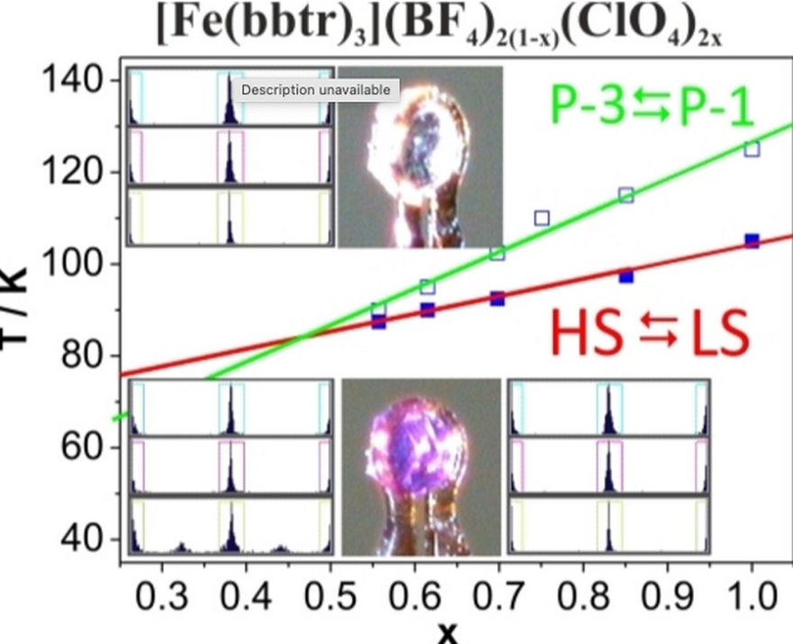
Spin crossover (SCO) occurs in octahedral coordination compounds containing d4-d7 metal ions, with a focus on Fe(II) coordination compounds. Spin crossover involves a reversible transition between high spin (HS) and low spin (LS) states, leading to significant changes in electronic and structural properties. Various factors such as temperature, pressure, light irradiation, and pulsed magnetic fields can trigger SCO. The change in spin state is associated with optical and dielectric properties, making these materials useful for applications like medical diagnostics, sensors, displays, memory devices, actuators and nanoscale integrated devices. Using the “settle mode” on a SQUID magnetometer the authors identify a thermally induced spin crossover, establishing the HS -> LS crossover centered at T1/2¯=76 K and T1/2=89 K. In [Fe(bbtr)3](ClO4)2 an isostructural perchlorate analogue of [Fe(bbtr)3](BF4)2, thermally induced spin crossover is preceded by a phase transition whereas in the (BF4)2 analogue the phase transition occurs at the start of the spin crossover. Variable pressure (<1 GPa), studies of the (ClO4)2 and (BF4)2 analogues indicate that this differentiation between the analogues only occurs in the thermal transition, while under pressure the SCO transition is followed by symmetry change in both complexes.
Citation: Ksiazek, M., Weselski, M., Kazmierczak, M., Polrolniczak, A., Katrusiak, A., Paliwoda, D., Kusz, J. & Bronisz, R. Extremely Slow Thermally-Induced Spin Crossover in the Two-Dimensional Network [Fe(bbtr)3](BF4)2. Chem. Eur. J., 15 (2023) 202302887. https://doi.org:10.1002/chem.202302887
ESS Contact: Damian Paliwoda
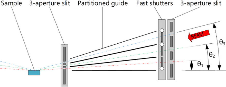
Despite their interest to many areas of chemistry, physics and biochemistry, the structural changes of surfaces and thin films at relevant timescales remain challenging to study. Time -of-flight neutron reflectometry can determine changes in surface composition and film thickness, but the speed of the measurements is limited by the available neutron flux and wavelengths. In a VR funded project, scientists and engineers from ESS, ISIS, and Uppsala University have collaborated to develop a novel fast shutter/aperture system which will allow dynamic characterization of surfaces and interfaces with sub-second time resolution on the FREIA instrument at ESS. This publication outlines a conceptual design and preliminary testing of this system that could also benefit the ZOOM SANS instrument at ISIS TS2.
For FREIA, the proposed design splits the incident beam is through three apertures, each with an individually controlled blocking paddle that can open and close the beam in between source pulses. For ZOOM, a fast aperture system is proposed with two slotted paddles which allow the aperture size to be modified during the source pulse meaning that different wavelengths observe a different aperture size. A simple prototype was designed and tested, using a single motor equipped with a paddle. The accuracy of the motors was tested in realistic vacuum conditions to ensure reproducible speeds (synching to the ISIS pulse), positional accuracy and positional stability. The paddle - an aluminum frame with boron carbide/epoxy composite cladding - successfully blocked the beam. The presented solution, which is reminiscent of a “neutron cat-flap", represents an effective and cost-efficient technical solution that could be useful to a wide range of neutron instrumentation applications.
Citation: Jacewicz, M., Elmer, J., Doutch, J., Arnold, T., Nightingale, J., Ekelof, T., Nylander, T. and Wacklin-Knecht, H. 'Newton' fast shutter system for neutron scattering instruments at the ESS and ISIS neutron sources. Nucl. Instrum. Methods Phys. Res. Sect. A-Accel. Spectrom. Dect. Assoc. Equip. 1055, doi:10.1016/j.nima.2023.168556 (2023).
ESS Contact: Tom Arnold
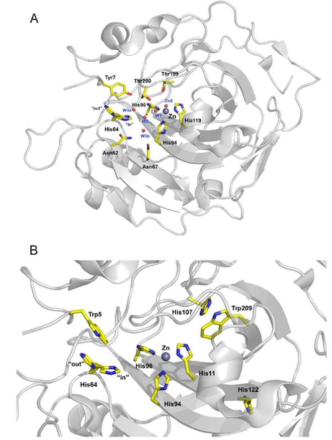
Human carbonic anhydrase II (HCA II) catalyses the reversible reaction of carbon dioxide and water to form bicarbonate and a proton. The catalytic step is performed by a zinc-bound hydroxide or water, while the rate limiting step is the proton transfer between the active site and the buffer or bulk solvent, which is mediated by a hydrogen-bonded network of water molecules and the proton shuttle residue His64. The work presented addresses the electrostatic and dynamic nature of His64, which displays two conformations in crystal structures; the inward pointing toward the zinc ion and the outward pointing toward the solvent.
Using NMR spectroscopy the pH titration curve for His64 was determined and can best be described by two pKa values: 6.25 and 7.60. These two pKa arise from its two conformations, “in” and “out”. His64 exists in both conformations at around 1:1 population, independent of pH. In addition, for both conformations the ratio of tautomers in their neutral form is also around 1:1. The life time of each conformation is short – on the ps to ns time scale. His64 displays high flexibility in both conformations – it is never static, but always ready to change conformation. The observed dynamic nature of His64 may be a prerequisite for the high enzymatic activity observed in HCA II.
Citation: Raum, H. N., Fisher, S. Z. & Weininger, U. Energetics and dynamics of the proton shuttle of carbonic anhydrase II. Cell. Mol. Life Sci. 80 (2023). doi.org:10.1007/s00018-023-04936-z
ESS Contact: Zoë Fisher
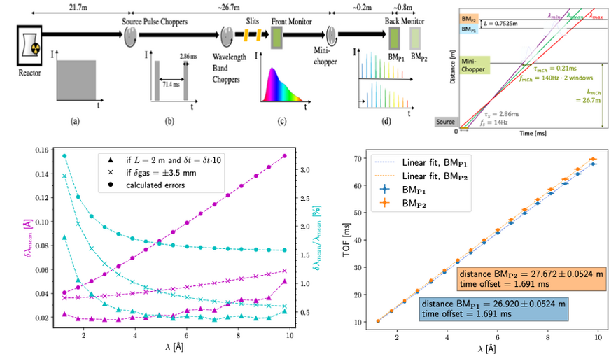
Determining the exact neutron flight path length in a real-world experimental setup can be challenging, due to the complex nature of neutron guide systems and the variations in the paths that individual neutrons might take. This publication presented a method for calibrating neutron scattering instruments, particularly focusing on the conversion of neutron time-of-flight (TOF) to neutron wavelength. This method employed a prototype mini-chopper and standard neutron monitors to measure the neutron flight path length from the neutron source to a monitor, leading to the determination of absolute wavelength with considerable precision.
Applying this method on a test beamline at ESS could open up several significant opportunities. The advantages of using a mini-chopper and standard neutron monitors include flexibility, simplicity, broad applicability, avoidance of sample-dependent uncertainties, and portability. When used on a test beamline, this method could enhance the accuracy of scientific experiments in neutron scattering instruments at spallation sources, ultimately contributing to the quality and reliability of research in neutron science.
Citation: Vergara, L., Arai, M., Woracek, R., Olsson, M., Quintanilla, A., Alcock, S., Nilsson, J., Kanaki, K., Kadletz, P. M., Kirstein, O., Hall-Wilton, R. & Tsapatsaris, N. Evaluation of a method for time-of-flight, wavelength and neutron flight path calibration for neutron scattering instruments by means of a mini-chopper and standard neutron monitors. J. Instrum. 18, P04012, doi:10.1088/1748-0221/18/04/P04012 (2023).
ESS Contact: Nikolaos Tsapatsiris
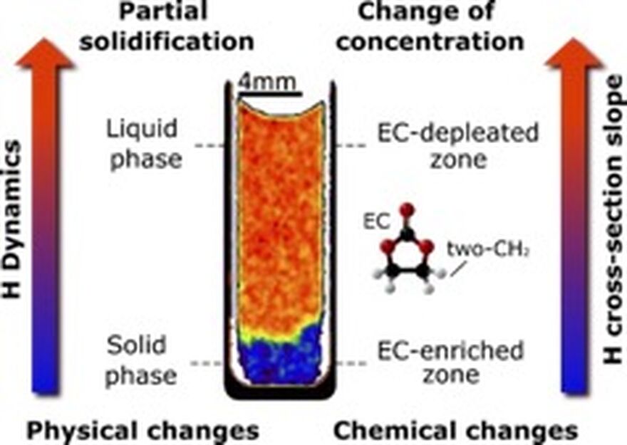
In recent years, batteries have become the most crucial technology for energy storage in portable and automotive applications. There is a strong necessity for improving performance under extreme conditions, e.g., sub-freezing temperatures and fast cycling. These conditions strongly affect the electrolyte, ultimately causing performance and safety issues. To study the behavior of the electrolyte operando without using contrast-enhancing isotopes, we developed a novel wavelength-resolved neutron imaging method capable of resolving H dynamics. This approach enables the identification of partial solidification and concentration changes in electrolytes due to temperature variations, presenting a new way to track real-time physical and chemical changes in H-containing compounds.
Spectroscopic neutron imaging (SNI) is a time-of-flight technique that spatially resolves spectroscopic features of hydrogenous molecules. This enables the identification of CHn functional groups based on their inelastic scattering properties, which are influenced by molecular vibrations and diffusion. Ongoing investigations have extended the application of the SNI to track aggregation state transitions in electrolytes, providing insights into the temperature limitations of solvents and mixtures. Furthermore, the method has been successfully applied to commercial batteries, correlating physico-chemical changes with performance loss. Recent applications also included temperature monitoring during welding of steel and the new method will be a powerful technique to enable first science at the ESS instrument ODIN.
This work was conducted at the ISIS pulsed neutron source and the ESS testbeamline V20 at Helmholtz Zentrum Berlin that operated between 2015 and 2019.
Citation:
Carreón Ruiz, E. R., Lee, J., Márquez Damián, J. I., Strobl, M., Burca, G., Woracek, R., Ebert, M.-O., Winter, E., Cochet, M., Höltschi, L., Kadletz, P. M., Zlobinski, M., Tremsin, A. S., Gubler, L., and Boillat, P. Spectroscopic neutron imaging for resolving hydrogen dynamics changes in battery electrolytes. Materials Today Advances, 19, 100405 (2023).
DOI: 10.1016/j.mtadv.2023.100405
ESS Contacts: Jose Ignacio Marquez Damian, Robin Woracek
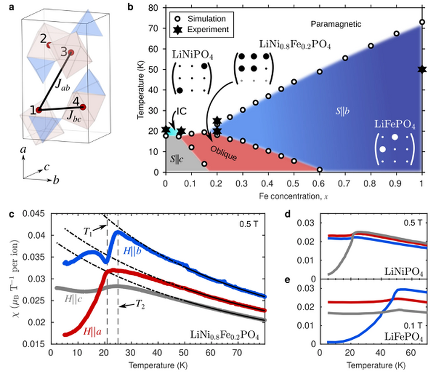
Control of magnetization and electric polarization is attractive in customising materials including those for data storage, sensors or antennae. In magnetoelectronic (ME) materials polarization can be controlled by a magnetic field and magnetization by an electric field, but the magnitude of the effect remains a challenge in the case of single-phase ME’s.
We have employed magnetic susceptibility and pyrocurrent measurements, neutron diffraction and Monte Carlo simulations to investigate the phase diagram of LiNi1−xFexPO4. This study demonstrates that the magnetoelectric properties are greatly influenced by substituting Ni2+ ions with Fe2+. Magnetoelectric couplings that are symmetry-forbidden in the parent compounds, LiNiPO4 and LiFePO4, are unlocked and the dominant coupling is enhanced by almost two orders of magnitude. The experimental and simulation data presented suggest that the observations have broader implications and that chemical tuning of oblique magnetoelectric phases represents a promising path for tailoring magnetoelectric material properties.
Citation: Fogh, E., Klemke, B., Reehuis, M., Bourges, P., Niedermayer, C., Holm-Dahlin, S., Zaharko, O., Schefer, J., Kristensen, A. B., Sørensen, M. K., Paeckel, S., Pedersen, K. S., Hansen, R. E., Pages, A., Moerner, K. K., Meucci, G., Soh, J.-R., Bombardi, A., Vaknin, D., Rønnow, H. M., Syljuåsen, O. F., Christensen, N. B. & Toft-Petersen, R. Tuning magnetoelectricity in a mixed-anisotropy antiferromagnet. Nat. Commun. 14, 3408, doi:10.1038/s41467-023-39128-7 (2023).
ESS Contact: Rasmus Toft-Petersen
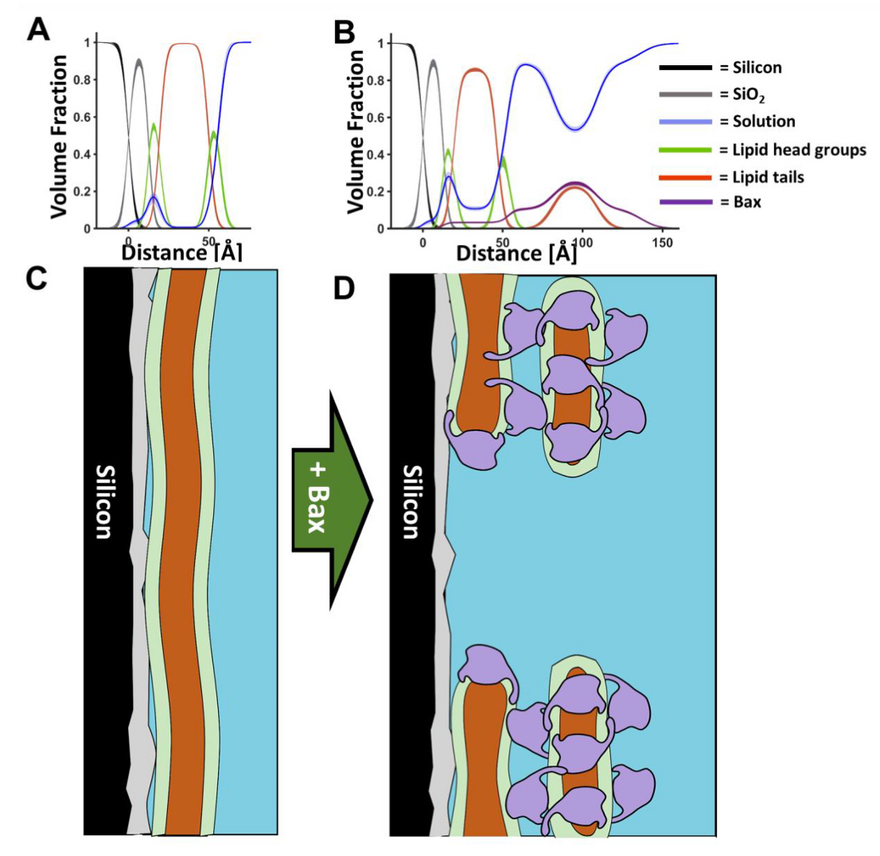
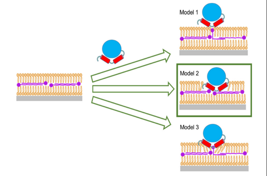
Apoptosis, or programmed cell death, is an essential process of life, tightly regulated by the Bcl-2 protein family with the pro-apoptotic Bax triggering cell death by perforating the mitochondrial outer membrane, unless it is captured by the pro-survival protein Bcl-2. Dysregulation of this process can cause a variety of pathological disorders including cancer. A research team from Umeå university, ESS and ISIS used time-resolved neutron reflectometry and Fourier-Transform infrared spectroscopy to identify two kinetically distinct phases in the Bax-membrane interaction corresponding to initial fast Bax adsorption to the membrane, followed by slower pore formation and the creation of Bax-lipid clusters on the membrane surface. These results give precise structural and kinetic details of how Bax remodels the mitochondrial outer membrane and reveal for the first time the presence of lipids in the Bax clusters associated with apoptotic pore formation.
Figure 1: A model for Bax pore formation. Component volume fraction profiles from before (A) and after (B) the interaction of Bax with mitochondrial model membranes. Below these are schematics of how this component distribution relates to the protein and lipid distributions across the surface.
Citation: Clifton, L.A., Wacklin-Knecht, H.P., Ådén, J., Mushtaq, A.U., Sparrman, T. and Gröbner, G. Creation of distinctive Bax-lipid complexes at mitochondrial membrane surfaces drives pore formation to initiate apoptosis, Science Advances 2023
ESS Contact: Hanna Wacklin-Knecht
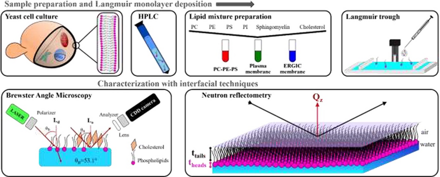
Here, the relationship between membrane composition and resultant structural features was investigated by surface pressure-area isotherms, Brewster angle microscopy and neutron reflectometry on in vitro membrane models of the mammalian plasma and endoplasmic-reticulum-Golgi intermediate compartment membranes. Natural extracted yeast lipids were used because, unlike synthetic lipids, the acyl chain saturation pattern of yeast and mammalian lipids are similar. The structural characterization of these membranes should yield insights into their function and malfunction.
To the authors' knowledge, this is the first work dedicated to fabricating Langmuir monolayers of natural lipids to mimic single membrane leaflets which have then been studied in situ by neutron reflectometry and Brewster angle microscopy. The development of such in vitro model systems could be exploited to study the interaction of membranes with novel molecules and colloids as well as bacterial or viral proteins.
Citation: Santamaria, A., Batchu, K. C., Fragneto, G., Laux, V., Haertlein, M., Darwish, T. A., Russell, R. A., Zaccai, N. R., Guzman, E. & Maestro, A. Investigation on the relationship between lipid composition and structure in model membranes composed of extracted natural phospholipids. J. Colloid Interface Sci. 637, 55-66, doi:10.1016/j.jcis.2023.01.043 (2023).
ESS Contact: Giovanna Fragneto
Despite being one of the most thoroughly characterised molecular crystals, hexamethylenetetramine (HMT) and its deuterated counterpart (DHMT) are still not fully understood, especially regarding anharmonic and nuclear quantum effects. To improve on that, this work involves an unprecedented combination of experimental techniques, including neutron and X-ray diffraction, inelastic neutron scattering, neutron transmission and Compton scattering.
The experimental results obtained for HMT and DHMT were benchmarked against the predictions of harmonic lattice calculation (HLD) simulations using CASTEP, and neutron scattering modelling using the NCrystal library. The experiments study the interplay between phonon anharmonicity and isotope and nuclear quantum effects related to the zero-point energies of photon and deuteron. The combined effects of the nuclear kinetic energy and anharmonicity of the local potentials of the mean force of the protons (deuterons) in the hydrogen bonds in HMT (DHMT), as well as the nuclear quantum delocalisation of both nuclear species are clearly visible in the systematic discrepancies between the HLD-based predictions for the nuclear momentum distribution (NMD) widths and the experimental values and the isotope effects on the NMD widths. Although these effects cannot be separated in the analysis of the NCS observables, it can be shown how they are intertwined, jointly dictating the nature of the nuclear quantum dynamics of protons and deuterons in both molecular crystals.
The study was carried out by researchers at the Rutherford Appleton Laboratory, in collaboration with researchers from Diamond Light Source, Tor Vergata University of Rome and the European Spallation Source.
Citation: Krzystyniak, M., Gutmann, M. J., Refson, K., Romanelli, G., Rudic, S., Capelli, S. C., Fortes, D., Magdysyuk, O., Márquez Damián, J. I. & Maciel-Pereira, G. Nuclear quantum dynamics in Hexamethylenetetramine and its deuterated counterpart: a DFT-augmented neutron study. Phys. Scr. 98, doi:10.1088/1402-4896/acb323 (2023).
ESS Contact: José Ignacio Márquez Damián
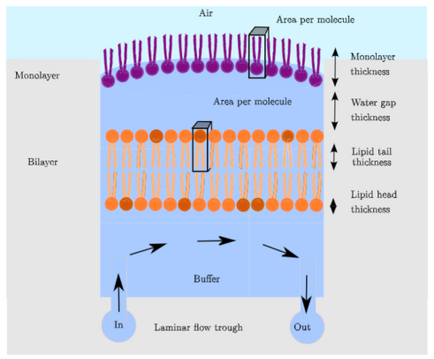
Membrane models comprised solely of lipids or lipid mixtures are an important tool for allowing detailed structural studies of membrane behaviour and interactions with relevant agents, such as proteins, peptides and DNA. In supported lipid bilayers the lipid bilayers can experience strong interactions (e.g. van-der-Waals attraction) with the solid surface, restricting the natural bilayer fluctuations and influencing lipid phase behaviour and dynamics. The lack of space between the bilayer and the substrate impedes the incorporation of membrane proteins. Here, we describe the development of a suspended bilayer approach, which removes the presence of a solid substrate.
A cationic surfactant (DODAB) monolayer is spread at the air–water interface and vesicles comprising a mixture of zwitterionic (POPC) and anionic (POPG) lipids are osmotically ruptured beneath the monolayer to form planar bilayers. Neutron reflectivity measurements have been used to show the complete rupture of the vesicles resulting in the formation of a continuous bilayer suspended 11 Å beneath the surfactant monolayer.
The suspended phospholipid bilayers can be probed by a variety of surface science techniques, such as X-ray reflectivity, Brewster angle microscopy, ellipsometry, reflection absorption infrared spectroscopy and neutron reflectivity, as well as providing the space to incorporate transmembrane proteins. The measured neutron reflectivity profiles are easy to interpret, as the interfacial region does not feature any of the superfluous layers such as silicon, silicon dioxide or gold that are typically present for floating bilayers formed on solid substrates.
Citation: Ayscough, S. E., Clifton, L. A., Skoda, M. W. A. & Titmuss, S. Suspended phospholipid bilayers: A new biological membrane mimetic. J. Colloid Interface Sci. 633, 1002-1011, doi:10.1016/j.jcis.2022.11.148 (2023).
ESS Contact: Sophie Ayscough
Science Highlights 2022
Laser powder-bed fusion (LPBF) is one of the most widely used Metal additive manufacturing (AM) technique, often referred to as 3D printing. In LPBF, a laser is used to selectively melt areas of a thin powder layer (powder-bed) according to a digital model. New layers are subsequently added and the process is repeated until the object is completed.
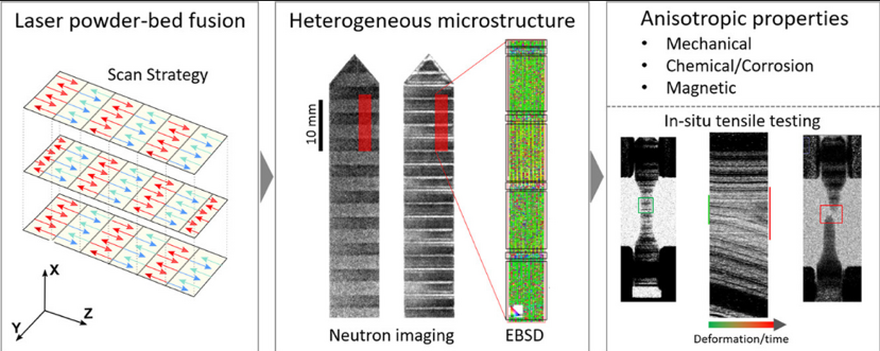
AM techniques are not only expanding our capabilities to design components with complex shapes, but also open new pathways for the creation of materials with tailored microstructures and hence mechanical as well as functional properties. The successful manufacturing of components by LPBF requires careful selection of process parameters (such as laser power, layer thickness, etc.) and scan strategies (laser beam movement). It has been known that the scan strategy can influence the microstructure and texture (i.e. the preferred orientation of the crystal grains within the material). Past studies have however largely overlooked that different scan strategies can inadvertently produce local microstructure alterations and preferential grain orientation over small areas – which can likely be attributed to a lack of suitable experimental techniques that work across cm-sized parts.
The current work reveals and explains this effect in parts made from 316L stainless steel, using a combination of neutron imaging, neutron diffraction and electron backscatter diffraction. During an in-situ tensile test, neutron imaging shows that – what seem to be minor changes in the scan strategy, can have significant effects on the mechanical performance and reveals locations where failure begins. The researchers also highlight that their manuscript can serve as an ideal reference for the – yet challenging – interpretation of neutron ‘Bragg edge imaging data’ in general, by showing the relationship between measured and simulated data, pole figures and EBSD in an unprecedented level of detail, e.g. benefitting future users of the future ODIN and BEER instruments.
Citation: Pacheco, V., Marattukalam, J. J., Karlsson, D., Dessieux, L., Van Tran, K., Beran, P., Manke, I., Kardjilov, N., Markötter, H., Sahlberg, M. & Woracek, R. On the relationship between laser scan strategy, texture variations and hidden nucleation sites for failure in laser powder-bed fusion. Materialia 26, 101614, doi:https://doi.org/10.1016/j.mtla.2022.101614 (2022).
ESS Contacts: Premek Beran, Robin Woracek
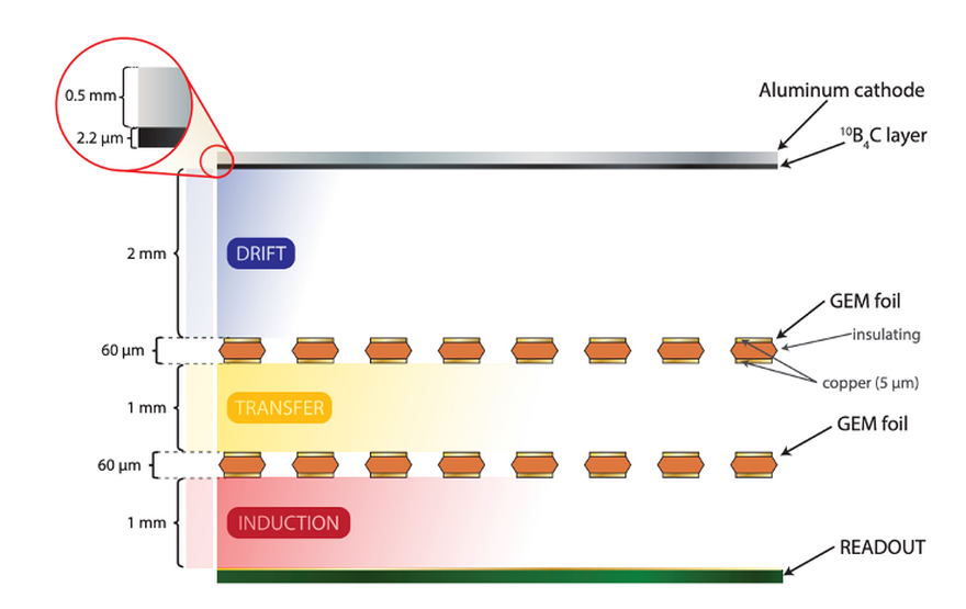
Advances in neutron scattering techniques in wide areas such as medicine and engineering are strongly dependent on detector developments with the capability to obtain bright monochromatic neutron sources. The 3He proportional counter neutron detector is widely used, with the advantages of a large neutron capture cross-section, low sensitivity to gamma-rays, and the possibility of working on a wide neutron energy range within the low energy region. Due to the shortage of 3He since the early 2000s, the science community works on the development of alternative technologies, employing 113Cd, 157Gd, 10B, and 6Li as the neutron converters instead of 3He. At Institute of Physics, University of Sao Paolo (IFUSP, Brazil), a double-GEM detector prototype, using a 10B4C layer as the neutron converter material, was developed and tested. The 10B4C neutron converting layer was coated on to a 0.5 mm Al cathode by ESS Detector Coatings Workshop at Linköping (Sweden). GEANT4 simulations were performed predicting an efficiency of (3.14 ± 0.10)%, agreeing within 2.7 ? with the experimental and analytic detection efficiencies obtained by the detector when tested in a 41.8 meV thermal neutron beam. The detector is position sensitive, equipped with a 256+256 strip readout connected to resistive chains, and achieves a spatial resolution better than 3 mm. The gain stability over time was also measured with a fluctuation of about 0.2% h−1 of the signal amplitude. A simple data acquisition with only 5 electronic channels is sufficient to operate this detector.
Citation: Serra, L. A., dos Santos, R. F., de Souza, G. G. A., Paulino, M. M. M., Souza, F. A., Moralles, M., da Luz, H. N., Bregant, M., Munhoz, M. G., Lai, C. C., Hoglund, C., Svensson, P. O., Robinson, L. & Hall-Wilton, R. Double-GEM based thermal neutron detector prototype. J. Instrum. 17, doi:10.1088/1748-0221/17/09/p09018 (2022).
ESS Contact: Chung-Chuan Lai, Per-Olof Svensson
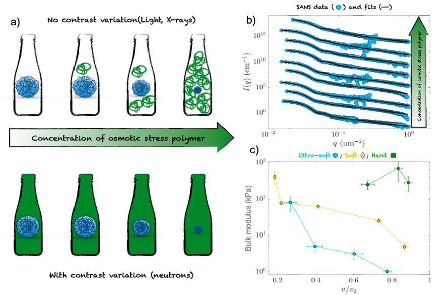
The bulk modulus, K, quantifies the elastic response of an object to an isotropic compression. For soft compressible colloids, knowing K is essential to accurately predict the suspension response to crowding. Most colloids have complex architectures characterized by different softness, which additionally depends on compression. Microgels and nanogels are one of the most commonly-used model systems to study the effects of particle compressibility on both phase transitions, crowding, and flow behavior. They often have a core region that is distinct from a surrounding fuzzy shell. This work determines the different values of K for the various morphological parts of individual nanogels and probes the change of K with compression. The method uses a partially deuterated polymer to exert the required isotropic stress and small-angle neutron scattering with contrast matching to determine the form factor of the particles without any scattering contribution from the polymer. We show a clear difference in softness, compressibility, and evolution of K between the shell of the nanogel and the rest of the particle, depending on the amount of cross-linker used in their synthesis.
Citation: Houston, J. E., Fruhner, L., de la Cotte, A., Gonzalez, J. R., Petrunin, A. V., Gasser, U., Schweins, R., Allgaier, J., Richtering, W., Fernandez-Nieves, A. & Scotti, A. Resolving the different bulk moduli within individual soft nanogels using small-angle neutron scattering. Sci. Adv. 8, doi:10.1126/sciadv.abn6129 (2022).
ESS Contact: Judith Houston
In 2022 the DTL1 Faraday cup was essential to the DTL1 commissioning, acting as a beam dump, measuring the proton current in real time and contributing to the first DTL1 acceptance scans. The device was designed at ESS in Lund (Sweden), and manufactured by RadiaBeam Technologies in Santa Monica (USA).
Before the DTL1 commissioning, MCNPX/ANSYS simulations were performed to optimize and validate the design of the DTL1 Faraday cup. In particular, thermo-mechanical simulations ensured a safe absorption and dissipation of the volumetric power-deposition due to 21 MeV protons and in the order of MW/cm3. As a result of the simulations, the operational limits and the proton-beam modes were defined to enable beam-dynamics studies with a proton current up to 65 mA, proton pulses up to 50 µs long, and repetition rate of either 1 or 14 Hz.
During the DTL1 commissioning, the DTL1 Faraday cup control system relied on EPICS for the control of the data acquisition, high-voltage, motion and water cooling system.
In preparation to the end of the DTL1 commissioning, activation and residual dose-rates were predicted in MCNPX/CINDER’90 calculations. The low residual dose-rate in the vicinity of the beam dump (below 2 µSv/h, after 8 hours of cooling time) allowed the relocation of the Faraday cup and its shielding the day after the DTL1 commissioning was over, thus quickly continuing the installation works for the other DTL sections.
Citation: E.M. Donegani, J. Cereijo-Garcia, S. Haghtalab, E. Laface, A. Olsson, L. Page, M. Ruelas, L. Zanini, Design and performance of the compact DTL1 Faraday cup for the high-power ESS NCL, Nuclear Inst. And Methods in Physics Research, A,doi: https://doi.org/10.1016/j.nima.2022.167827 (2022).
ESS Contact: Elena Donegani
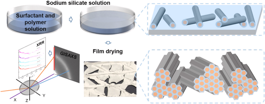
Ordered mesoporous silica materials in thin-film geometry are in demand due to their applications in chemical sensors and separation. An environmentally friendly and inexpensive silica source, sodium silicate solution, was applied to synthesize a free-standing film with a wormlike structure formed at the air/liquid interface. This reorganized into a 2D hexagonal ordered structure while drying at room temperature, after removal from the air/solution interface. Small angle X-ray scattering (SAXS) data show the effect of the composition of the solution used for film formation on the mesostructure of the silica films. Variations in the silica concentration in solution directly affect silica incorporation into the film and the degree of mesostructural ordering in the dry film. The initial film formation time was estimated by the change in surface pressure. The film with best ordering but also maximum initial film formation time, ~13h, was achieved with a concentration of 50-60 mM SiO2. Data from in situ grazing incidence small angle X-ray scattering (GISAXS) and X-ray reflectivity (XRR) were used to propose a possible formation process for the mesostructured silica film.
Citation: Di, A. D., Schmitt, J., Elstone, N., Arnold, T. & Edler, K. J. In situ X-ray reflectivity and GISAXS study of mesoporous silica films grown from sodium silicate solution precursors. Microporous Mesoporous Mat. 341, doi:10.1016/j.micromeso.2022.112018 (2022).
ESS Contact: Tom Arnold
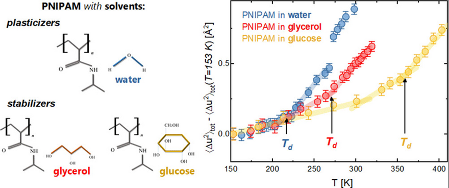
With a repeating unit including both hydrophilic amide and hydrophobic isopropyl groups, poly-N-isopropylacrylamide (PNIPAM) is a paradigm example of a smart polymer. PNIPAM has significance as a simple but predictive protein model, since the coil-to-globule transition in many aspects resembles the cold renaturation of small globular proteins. Recently, neutron scattering experiments and molecular dynamics simulations identified the existence of a dynamical transition in weakly hydrated PNIPAM samples at a critical temperature Td ≈ 220 K. Common features of the transition, manifested by both biopolymers and PNIPAM, are (i) the intimate coupling between solvent and solute dynamics, and (ii) the connection with the inherent complexity of the polymer repeating unit. This work studies how the addition of stabilizing agents, like sugars (glucose) and polyols (glycerol), impacts the dynamical transition of hydrated PNIPAM chains. When the polymer is embedded in glycerol or glucose, the dynamical transition occurs even in the absence of water, although an upshift of the transition temperature Td is observed. In the presence of binary solvent mixtures, the competition between water and cosolvent in interaction with PNIPAM results in a strong dependence of Td on the water content. The comparison with proteins reveals the existence of strong analogies between PNIPAM and protein behavior, not only in purely hydrated conditions but even in the presence of more complex solvent environments.
Citation: Rosi, B. P., D'Angelo, A., Buratti, E., Zanatta, M., Tavagnacco, L., Natali, F., Zamponi, M., Noferini, D., Corezzi, S., Zaccarelli, E., Comez, L., Sacchetti, F., Paciaroni, A., Petrillo, C. & Orecchini, A. Impact of the Environment on the PNIPAM Dynamical Transition Probed by Elastic Neutron Scattering. Macromolecules 55, 4752-4765, doi:10.1021/acs.macromol.2c00177 (2022).
ESS Contact: Daria Noferini
Residual stress (and thereby elastic strain) is the stress that remains in a sample when no external forces are applied. These internal stresses add to those arising from externally applied loads and, if they are not detected, they can give rise to unexpected behaviours and premature failure. A well-established technique used for strain measurements is based on neutron diffraction (or Bragg scattering). Measurement of the position of Bragg peaks from diffraction allows the determination of lattice spacings, while the measurement of the relative shift in the positions provides information on lattice strains.
Bragg edge strain tomography seeks to determine the spatial distribution of strain inside a polycrystalline sample from the change in the neutron transmission spectra near a Bragg edge. Bragg edges can be considered as crystallographic fingerprints whose locations and shapes depend on microstructure and strain distribution. In the current approach, the Bragg edge strain tomography requires extraction of the second moment (variance about zero) of the strain distribution. This paper is the first experimentally proof that the second moment can be reliably measured for a previously well characterized sample.
Citation: Fogarty, K., Ametova, E., Burca, G., Korsunsky, A. M., Schmidt, S., Withers, P. J. & Lionheart, W. R. B. Recovering the second moment of the strain distribution from neutron Bragg edge data. Appl. Phys. Lett. 120, doi:10.1063/5.0085896 (2022)
ESS Contact: Søren Schmidt
The development of high-entropy alloys (HEA) has grown rapidly since their discovery. In HEAs, solid solutions with simple crystal structures, e.g. body-centred cubic (bcc), cubic close-packing (fcc) or hexagonal close-packing (hcp), are assumed to form due to the high configurational entropy from the mixture of at least five different elements in near equiatomic concentrations. Depending on the composition, these alloys may possess desirable properties such as high strength at various temperatures, high hardness, high ductility, and excellent wear resistance. Good oxidation resistance and promising mechanical properties make the AlCoCrFeNi alloy one of the most studied HEAs and a candidate for high-temperature applications. The aim of this study is to increase the understanding of the microstructure and phase formation of gas atomised AlCoCrFeNi with different particle sizes. A combination of X-ray and neutron diffraction with electron microscopy was utilised to determine the crystal structure, with emphasis on the atomic occupations in the as-atomised powder state. Moreover, in-situ synchrotron X-ray and neutron diffraction experiments revealed the phase transformation across an important temperature range.
The results show that the powder crystallises in an ordered cubic B2 (CsCl-type) structure with the (0 0 0) position preferentially occupied by Co and Ni and the (½ ½ ½) position preferentially occupied by Al and Fe. And Cr is equally distributed between both positions. During heat-treatment of the powder, the B2 phase decomposes into fcc and σ phases, and the final phase composition is highly dependent on the heating rate. Finally, it was demonstrated that the pre-annealing of the atomised powder influences the microstructure of sintered parts, with a lower amount of σ phase in the final component if a pre-annealing step of the powder is employed. Hence, it can be concluded that the microstructure can be designed by proper selection of the feedstock material used for manufacturing.
Cite: Karlsson, D., Beran, P., Riekehr, L., Tseng, J. C., Harlin, P., Jansson, U. & Cedervall, J. Structure and phase transformations in gas atomized AlCoCrFeNi high entropy alloy powders. J. Alloy. Compd. 893, 8, doi:10.1016/j.jallcom.2021.162060 (2022).
ESS contact: Premek Beran
Some of the most well-known magnetic materials can be surprising complex. A good example is magnetite, discovered by the ancient Greeks. Magnetite has a so-called inverse spinel structure, where the magnetic moment resides on two distinct sublattices, which can give rise to complex magnetisation curves when the temperature is lowered. This work studies another inverse spinel compound, Co2VO4, where the magnetic susceptibility goes to zero and even reverses sign when the temperature is lowered. By examining the temperature evolution of magnetic structures using neutron diffraction, magnetisation, polarised muon spectroscopy, and performing electronic structure calculations, the team established the existence of several phase transitions. Neutron diffraction revealed the structure to be composed of two nearly balanced antiparallel FM sublattices, on the so-called A and B sites, giving rise to a net ferrimagnetic phase that evolves with temperature. A significant difference between the temperature evolution of the ordered moment of the two sublattices causes a tipping of this balance, giving rise to a change in sign of the net magnetic moment in the unit cell. This temperature evolution is accompanied by anomalous magnetic dynamics well below the ordering temperature, as revealed by muon spectroscopy. One interpretation of these results is the existence of a mechnanism also proposed for magnetite, namely that some of the electrons responsible for magnetism on one of the sublattices are delocalized at high temperatures, but gradually localise as the temperature is lowered. This serve to change both the net magnetic moments and the interactions between them, giving rise to the magnetisation reversal. Density functional theory (DFT) calculations were carried out, supporting this interpretation further. Using these modern experimental and computational techniques, the team have shed more light on the underlying mechanisms behind the bulk properties of this otherwise well-studied material.
Cite: Kademane, A. B., Bhandari, C., Paudyal, D., Cottrell, S., Das, P., Liu, Y., Yiu, Y., Kumar, C. M. N., Siemensmeyer, K., Hoser, A., Quintero-Castro, D. L., Vaknin, D. & Toft-Petersen, R. Magnetization reversal driven by electron localization-delocalization crossover in the inverse spinel Co2VO4. Phys. Rev. B 105, 9, doi:10.1103/PhysRevB.105.094408 (2022).
ESS Contact: Rasmus Toft-Petersen
Published 4 May 2022: Spin liquids are a topical and fascinating subject of condensed matter physics. Their cooking recipe is a combination of antiferromagnetic interaction with strong frustration, which leads to a rich variety of highly correlated spin states rather than to conventional order. Our experiment shows the first observation of a chiral spin liquid state. Our discovery has far-reaching implications; within the fundamental theory of magnetic ordering, it is argued that chirality can twist the spins into an inextricable structure of short-range entities that constitute a classical ground state. The lack of periodic order is contrary to the common expectation in condensed matter physics and thought to be possible only by quantum effects. As a side note, the identified scenario has a commonality with the possible behavior of subnuclear matter.
Cite: Schweika, W., Valldor, M., Reim, J.D., Rößler, U.K. Chiral Spin Liquid Ground State in YBaCo3FeO7. Phys. Rev. X (2022).
ESS contact: Werner Schweika
Link: https://journals.aps.org/prx/accepted/2e07eKfbX2119b0786c707101fd7facf0e840e4bf
Published 23 February 2022: The fourth enzymatic reaction in de novo pyrimidine biosynthesis, the oxidation of dihydroorotate to orotate, is catalyzed by dihydroorotate dehydrogenase (DHODH). Enzymes belonging to the DHODH Class II are membrane-bound proteins that use ubiquinones as their electron acceptors. This study uses neutron reflectometry (NR) to investigate the interaction between N-terminally truncated human DHODH (HsΔ29DHODH) or Escherichia coli DHODH (EcDHODH) with ubiquinone (Q10) in a supported lipid membrane. The benefit of using NR for this work is that one can inspect in situ and in solution the mechanism of enzyme binding to the lipid membranes. Additionally, it was possible to determine the location of the Q10 by using chain deuterated d63-POPC lipids provided by ESS through the Deuteration and Macromolecular Xtallisation platform (DEMAX) pilot proposal call scheme launched in 2019. This work has determined that the Q10 is at the centre of the lipid bilayers and that upon binding both DHODH enzymes penetrate into the outer membrane leaflet towards the Q10. This interaction had an enzyme-dependent binding strength for different lipid bilayer compositions and the presence/absence of Q10. This is the first time the membrane penetration has been observed for DHODH enzymes lacking a N-terminal transmembrane anchor.
Cite: Orozco Rodriguez, J. M., Wacklin-Knecht, H. P., Clifton, L. A., Bogojevic, O., Leung, A., Fragneto, G. & Knecht, W. New Insights into the Interaction of Class II Dihydroorotate Dehydrogenases with Ubiquinone in Lipid Bilayers as a Function of Lipid Composition. Int. J. Mol. Sci. 23, 2437 (2022).
ESS contact: Hanna Wacklin-Knecht
Link: https://www.mdpi.com/1422-0067/23/5/2437
Science Highlights 2021
Published 17 December 2021: The cold and thermal neutrons produced at the European Spallation Source are guided to the sample locations of the instruments, some of which are up to around 150 m distant from the target, by neutron guides. These guides are coated with supermirrors, which consist of alternating layers of, for example, Ni and Ti, and have differing performance depending on their properties. The neutrons which are not reflected, may instead be absorbed within the supermirror coating, resulting in the emission of gamma-rays. The shielding to minimise the impact of gamma-rays, both for radiation safety and for instrument performance, is a major component of each instrument. While there are theoretical and computational models describing the absorption levels in supermirrors, there has so far not been an experimental validation carried out previously. Thus, experiments were carried out at the SuperADAM instrument at the Insitut Laue-Langevin to measure the gamma-ray production from two types of supermirrors when illuminated with neutrons. The results obtained provide a valuable validation for theoretical and computational models and can be used as input to Monte-Carlo calculations for the design of neutron instrument shielding.
Cite: DiJulio D.D. et al., Measurements of the neutron absorption in supermirror coatings. Nuclear Instruments and Methods in Physics Research, A 1025(2022) 166088 doi:10.1016/j.nima.2021.166088
ESS contacts: Douglas DiJulio, Valentina Santoro, Günter Muhrer
Link: https://www.sciencedirect.com/science/article/pii/S0168900221010019
(Image: Reprinted from D.D. DiJulio et al., Measurements of the neutron absorption in supermirror coatings. Nuclear Instruments and Methods in Physics Research, A 1025(2022) 166088 with permission from Elsevier)
Published 12 November 2021: Superconductivity was first discovered over 100 years ago, but there are still many open questions. While conventional superconductors are well explained by the BCS theory, a large number of ‘unconventional superconductors’ behave differently. One example is CeCu2Si2 – the first heavy fermion superconductor to be discovered in 1979. But what does heavy fermion mean here?
In a superconductor, the charge carriers (usually electrons) pair up to form a Cooper pair. In a heavy fermion material, the charge carriers behave as though they have a mass many times that of a free electron, pushing all the energy scales in the materials lower. BCS theory cannot be applied to materials like this, and so a lot of questions remain over the superconducting state. To study this, we looked at the flux-line lattice that develops inside CeCu2Si2 when it is put in a magnetic field. Normally, a superconductor expels magnetic field, but many superconductors, like CeCu2Si2, allow magnetic flux to enter as small tubes, or flux lines, where the material is not superconducting. The rest of the material then stays superconducting up to larger fields, or with larger applied currents. We have used small-angle neutron scattering to diffract off the flux-line lattice, as the size and arrangement of the flux lines tells us about the underlying superconductivity. An example diffraction pattern is shown below, before and after processing via Bayesian methods to extract more information from the scattered neutrons. CeCu2Si2 shows a very unusual increase in flux-line intensity towards the edge of the superconducting phase, whereas in most other superconductors, this intensity drops towards zero, indicating that the superconductivity is not destroyed in the same way as in almost all other superconductors.
Cite: Campillo, E. et al., Observations of the effect of strong Pauli paramagnetism on the vortex lattice in superconducting CeCu2Si2. Phys. Rev. B, 104, 184508, 2021, doi: 10.1103/PhysRevB.104.184508
ESS contact: Alex Holmes
Link: https://journals.aps.org/prb/abstract/10.1103/PhysRevB.104.184508
Published 1 November 2021: The use of scintillators and cameras is extremely widespread in the field of neutron radiography, both for its cost-effectiveness and for its simplicity. One drawback of such an approach is that the scintillator itself is the limiting factor in terms of achievable resolution and detection efficiency. Furthermore, this configuration offers very little in terms of time resolution and therefore can be used for a limited number of very specific Time-of-Flight applications. Recent developments in event-driven detectors allow the recording and reconstruction of the shape of light spots emitted by the scintillator after a single particle interaction, allowing reconstruction of the Center-of-Mass of the interaction within the scintillator. This enables a more precise localisation of the single event and provides a pathway to overcome the scintillator thickness limitation and increase the effective spatial resolution of the system, while still retaining the ease of use and flexibility of traditional camera systems. The detection of single events allows also to retain the timing information, making this approach suitable for Time-of-Flight applications.
In the paper, such a detector is presented, and it is shown that it is capable of Time-of-Flight imaging with an adjustable field-of-view, ad-hoc binning and re-binning of data based on the requirements of the experiment and the possibility of particle discrimination via the analysis of the event shape in space and time. We believe that this novel concept might replace regular cameras in neutron imaging detectors as it provides superior detection capabilities. We demonstrate an increase by a factor 3 in image resolution and an increase by up to a factor of 7.5 in signal-to-noise for thermal neutron imaging. Such a detector is not limited only to imaging, and new approaches to detection for diffraction experiments are also a clear outlook.
Cite: Losko, A.S., New perspectives for neutron imaging through advanced event-mode data acquisition. Sci Rep, 11, 21360, 2021, doi: 10.1038/s41598-021-00822-5
ESS contact: Manuel Morgano
Link: https://www.nature.com/articles/s41598-021-00822-5
Published 6 October 2021: In March 2020, the Deuteration and Macromolecular Crystallisation (DEMAX) groups at the ESS issued a special call for proposals related to Covid-19 research. One of the projects has led to a larger collaboration between DEMAX, Frank Kozielski (University College London), and Wolfgang Knecht (Lund Protein Production Platform, LP3, at Lund University) and now the FragMAX platform of the MAX IV laboratory. The project is to find small molecule inhibitors that can block the SARS CoV-2 replication machinery. Non-structural protein 10 (nsp10) is a conserved stimulator of two enzymes crucial for viral replication, nsp14 and nsp16, exhibiting exoribonuclease and methlytransferase activities. Interfering with RNA proofreading or RNA cap formation represents intervention strategies that can be exploited to inhibit viral replication. Fragment-based screening using nano differential scanning fluorometry, x-ray crystallography and hit confirmation by microscale thermophoresis was carried out to identify ligands and measure their binding affinities to SARS-CoV-2 nsp10. Four fragments were identified that were found to bind to two distinct binding sites on nsp10: one can be modelled to where it would be located in the nsp14-nsp10 interface (Figure 1) and the other to the protein-protein interace of the nsp16-nsp10 complex. These fragments now serve as starting points for the development of more potent analogues using fragment growing techniques and structure-based drug design. This research used unique capabilities found at FragMAX and the BioMAX beamline, accessing a fragment library and making use of prioritized access to the BioMAX beamline, all during the ongoing pandemic
Cite: Kozielski F. et al. Identification of fragments binding to SARS-CoV-2 nsp10 reveals ligand-binding sites in conserved interfaces between nsp10 and nsp14/16. RSC Chemical Biology, 2021, doi:10.1039/D1CB00135C
ESS Contact: Zoë Fisher; LP3 Contact: Wolfgang Knecht; UCL Contact: Frank Kozielski; FragMAX Contact: Tobias Krojer
Link: https://pubs.rsc.org/en/content/articlelanding/2021/cb/d1cb00135
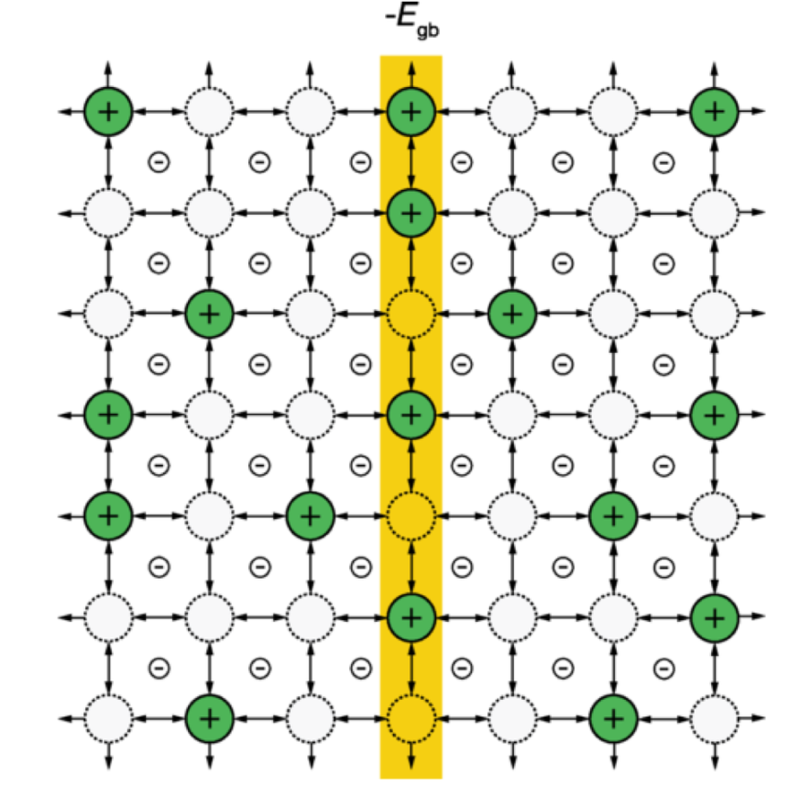
Published 24 September 2021: Solid electrolytes are ionic conductors with potential applications in next generation batteries. However, prediction of the equilibrium distribution of ions within these materials, which are crystalline solids with grain broundaries and heterointerfaces, is a long-standing problem in solid-state physics. Near to these interface regions, the concentration of individual ionic species can deviate significantly from their formal bulk values, giving rise to so-called “space-charge” regions. Using kinetic Monte Carlo simulations of a three-dimensional Coulomb lattice gas, this work has show that these space-charge regions may exhibit features similar to liquid-state materials, such as ionic liquids and concentrated electrolytes. The analysis of this work utilised Bayesian inference to compare traditional solid state dilute limit models with more complex oscillatory models.
Cite: Dean, J.M. et al. Overscreening and Underscreening in Solid-Electrolyte Grain Boundary Space-Charge Layers. Phys. Rev. Lett., 2021, 127, 135502, doi: 10.1103/PhysRevLett.127.135502
ESS Contact: Andrew McCluskey
Link: https://journals.aps.org/prl/abstract/10.1103/PhysRevLett.127.135502
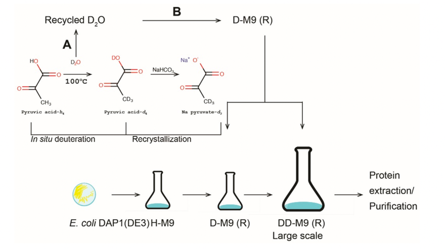
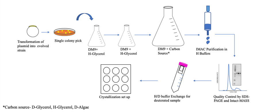
Published 7 September 2021: Labelling of proteins with deuterium (2H) is often necessary for structural biology techniques, such as neutron crystallography, NMR spectroscopy, and small-angle neutron scattering. However, perdeuteration in which all protium (1H) atoms are replaced by deuterium is a costly process. Typically, an expression host such as Escherichia coli is grown in a defined medium (M9) with heavy water as the solvent and a deuterated carbon source such as glucose-d7 or glycerol-d8. An alternative carbon source, pyruvate, can be deuterated in-house relatively simply, but the growth of E. coli on this carbon source is very poor. To circumvent this problem, adaptive laboratory evolution was used to obtain E. coli strains with a greatly improved growth rate on pyruvate, and using pyruvate-d3 as carbon source, >90% deuterated triose-phosphate isomerase was produced in sufficient quantities for large volume crystal production and subsequent analysis by neutron crystallography.
Cite: Kelpšas, V. et al. Evolving Escherichia coli Host Strains for Efficient Deuterium Labeling of Recombinant Proteins Using Sodium Pyruvate-d3. Int. J. Mol. Sci., 2021, 22, 9678, doi: 10.3390/ijms22189678
ESS Contact: Anna Leung
Link: https://www.mdpi.com/1422-0067/22/18/9678
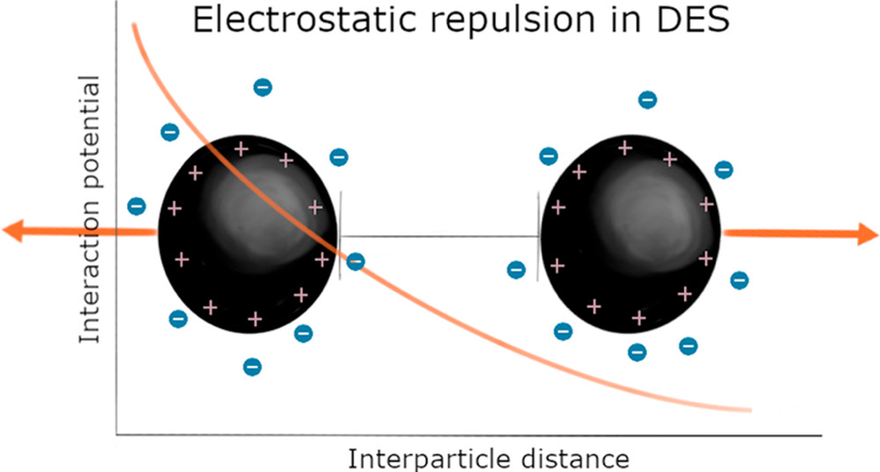
Published 30 August 2021: Electrostatic interactions play an essential role in biological and technological processes. From ion motion in batteries to protein function in living cells, charge modulation dictates the function of processes that involve ions. The traditional consensus dictates that high ion concentrations lead to negligible long-range electrostatic interactions. This research demonstrates that electrostatic correlations prevail in deep eutectic solvents where intrinsic ion concentrations often surpass 2.5 M. By investigating intermicellar interactions in 1:2 choline chloride:glycerol and 1:2 choline bromide:glycerol using small-angle neutron scattering, long-range electrostatic repulsions between charged colloidal particles were observed. Experiments were performed on Sans2d (ISIS Pulsed Neutron Source) and D11 (Institute Laue-Langevin).
Cite: Sanchez-Fernandez, A. et al. Long-Range Electrostatic Colloidal Interactions and Specific Ion Effects in Deep Eutectic Solvents. J. Am. Chem. Soc., 2021, 143, 35, 14158-14168 doi: doi.org/10.1021/jacs.1c04781
ESS contact: Andrew Jackson
Link: https://pubs.acs.org/doi/10.1021/jacs.1c04781
Published 17 August 2021: Hydrogen (1H) atoms form the basis of the hydrogen bond that is not scattered by X-ray crystallography due to its poor scattering power. Neutron protein crystallography (NPX) is a powerful tool that is capable of locating hydrogens and study the significance of hydrogen bonding interactions in biomacromolecules. However, due to the requirement for large crystals very few neutron structures have been deposited in PDB with no membrane protein structure determined yet. Additionally, 1H has a negative scattering length and large incoherent cross-section giving rise to a significant background noise in neutron data collection. This effect can be minimised by isotopic substitution of 1H with its heavier isotope deuterium (2H or D) leading to less ambiguous data analysis and better structure interpretation. Overall ~25% H atoms in a protein are solvent can be exchanged by dissolving in heavy water. However, complete deuterium labelling (perdeuteration) is required for the remaining 75% H atoms. In this work, an optimised methodology for large scale production of perdeuterated bacterial outer membrane protein F (OmpF) has been designed. OmpF was produced in deuterated minimal medium with different carbon sources. Mass spectrometry and thermal stability experiments verified the purity and level of deuteration of OmpF protein. We obtained protiated and perdeuterated OmpF crystals that diffracted X-rays to a resolution of 1.9 Å. No significant effect of deuterium-labelling was observed on structural properties of protein. This work lays the foundation for structural studies of membrane protein by neutron diffraction in future.
Cite: Aggarwal, S. A protocol for production of perdeuterated OmpF porin for neutron crystallography. Protein Expression and Purification, 188, 105954, 2021, doi: 10.1016/j.pep.2021.105954
ESS contact: Esko Oksanen, Zoë Fisher; Lund University contact: Claes von Wachenfeldt
Link: https://www.sciencedirect.com/science/article/pii/S1046592821001376?via%3Dihub

Published 13 August 2021: Quintessential frustrated magnetic materials are those based on a triangular hyperkagome crystalline lattice and include the isostructural compounds Gd3Ga5O12 (GGG) and Yb3Ga5O12 (YGG). Our previous studies (Science 350, 6257 (2015)) uncovered a most unusual long range emergent director state in GGG derived from magnetic spins arranged on a ten-ion loop despite a disordered state at the local level. In this most recent work we show that the director state is also found in YGG, albeit correlated over a much reduced distance, revealing the ubiquity of this unusual state of matter. Neutron scattering experiments were performed on single crystal YGG, using the polarised diffuse scattering spectrometer, D7, at the ILL, the cold neutron scattering spectrometer, CNCS at the SNS and the thermal chopper spectrometer, IN4 at the ILL. An understanding of these novel states of matter in magnetic materials will result in technological breakthroughs beneficial to our spintronic society.
PHD students: R.Edberg (KTH), L: Sandberg (KU-ESS) and I.M. Bakke (Oslo University-ESS), supervised by Pascale Deen, Senior Scientist for Spectroscopy at ESS, Pascale Deen, and Adjunct Associate Professor, Niels Bohr Institute , University of Copenhagen.
Cite: Sandberg, L. Ø. et al. Emergent behavior in the frustrated Yb3Ga5O12 garnet. Phys. Rev. B. 104, 064425, doi:10.1103/PhysRevB.104.064425 (2021).
ESS Contact: Pascale Deen
Link: https://journals.aps.org/prb/abstract/10.1103/PhysRevB.104.064425
Published 29 July 2021: The H- ion is often used for the accumulation of high-intensity proton populations in storage rings. This is done by stripping both its electrons (using either thin intercepting foils or laser stimulation) and bringing them into orbit with a circulating proton bunch. In doing so, higher intensities can be extracted from a ring than would be possible by simply injecting protons. The difficulty is reaching the ring: H- tends to have its weakly bound outer electron easily stripped away prior to the injection stage. This produces a neutral hydrogen atom, which follows a drift trajectory to ultimately collide and deposit most of its energy into external components, causing unwanted induced radioactivity. In this work, we summarize the theory governing the various types of stripping, from interaction with external fields or blackbody radiation, to collisions with adjacent H- ions. Simulated results are shown for practical test cases, where we demonstrate that the large majority of stripped hydrogen atoms deposit their energy into a final line-of-sight beam dump at the end of a linac or straight section, potentially relaxing the concerns for induced radioactivity of beam-line components.
Cite: Folsom B.T. et al. Stripping mechanisms and remediation for H- beams. Phys Rev Accel Beams 24, 074201, doi: 10.1103/PhysRevAccelBeams.24.074201
ESS Contact: Ben Folsom
Link: paper - https://link.aps.org/doi/10.1103/PhysRevAccelBeams.24.074201
Link: project video - https://www.youtube.com/watch?v=qAnvft0nAlg
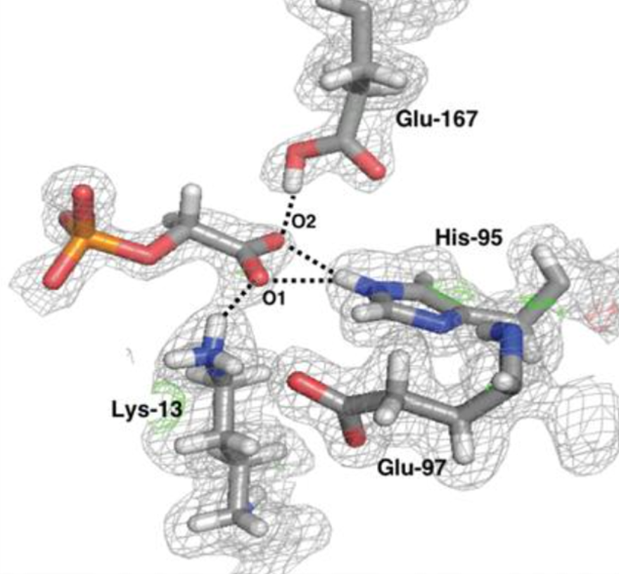
Published 3 June 2021: Triosephosphate isomerase (TIM) is a key enzyme in glycolysis that catalyses the interconversion of glyceraldehyde 3-phosphate and dihydroxyacetone phosphate. This simple reaction involves the shuttling of protons mediated by protolysable side chains. It is an example of the case of an enzyme mechanism where a long-standing controversy between competing models of proton movements can be resolved by neutron crystallography. The structures presented show that with the transition state analogue 2-phosphoglycolate (PGA) the postulated general base Glu167 is protonated, confirming Glu167 as the general base. The deuteron is clearly localised on Glu167, suggesting an asymmetric hydrogen bond instead of a low-barrier hydrogen bond. The full picture of the active-site protonation states allowed an investigation of the reaction mechanism using density-functional theory calculations.
Cite: Kelpšas V. et al. Neutron structures of Leishmania mexicana triosephosphate isomerase in complex with reaction-intermediate mimics shed light on the proton-shuttling steps. IUCrJ, 8(4), 633-643. 2021. doi:10.1107/S2052252521004619
ESS Contact: Esko Oksanen
Link: Paper - https://doi.org/10.1107/S2052252521004619
Link: Commentary - https://doi.org/10.1107/S205225252100573X